
Office Phone Number: (832) 644-8930
New Patient Appointments: (346) 662-1220
Cardiovascular diseases are among the leading causes of morbidity and mortality worldwide. Early and accurate diagnosis is crucial for effective management and treatment of these conditions. One of the most valuable tools in the early detection of vascular diseases is Doppler ultrasound. This non-invasive imaging technique allows doctors to assess blood flow and identify potential issues within the vascular system. At Modern Heart and Vascular, a cardiovascular and preventative medicine practice with several offices in the Houston area, Doppler ultrasound plays a vital role in the diagnostic process.
Doppler ultrasound employs high-frequency sound waves to create detailed images of the blood vessels and measure the velocity of blood flow. This information helps healthcare professionals diagnose a range of vascular conditions, including arterial blockages, blood clots, and abnormalities in blood flow. By providing real-time data, Doppler ultrasound enables prompt and accurate evaluations, ensuring that patients receive the necessary interventions without delay.
Furthermore, Doppler ultrasound is a safe and painless procedure with no exposure to ionizing radiation, making it suitable for repeated use. This allows for ongoing monitoring and assessment of vascular health, particularly in patients with chronic conditions such as peripheral arterial disease and carotid artery disease.
In this article, we will explore the role of Doppler ultrasound in diagnosing vascular diseases, highlighting its significance, applications, benefits, and the expertise provided by Modern Heart and Vascular. Understanding how this technology works and its application in cardiovascular medicine can help patients better appreciate its importance in their healthcare journey.
Doppler ultrasound is a type of imaging test that uses sound waves to evaluate the movement of blood through blood vessels. It is named after the Doppler effect, which is the change in frequency of sound or light waves as they move relative to an observer. This technology helps doctors see whether blood flow in the arteries and veins is normal or if there are blockages or other problems.
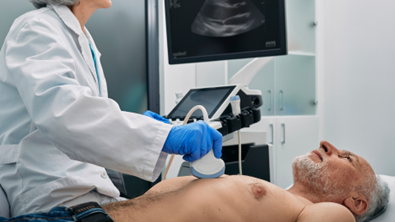
During a Doppler ultrasound, a handheld device called a transducer sends high-frequency sound waves into the body. These waves bounce off moving blood cells, and the echoes are captured by the transducer. The computer connected to the transducer converts these echoes into visual and audible data, providing real-time images and sounds of blood flow. This non-invasive test offers a clear view of blood circulation without the need for needles or incisions.
Importance of Doppler Ultrasound in Vascular Disease Diagnosis
The timely diagnosis of vascular diseases is crucial for effective treatment. Doppler ultrasound plays an important role in identifying issues such as blood clots, blockages, and abnormal blood flow. This technology allows for early detection, which can prevent serious complications like heart attacks, strokes, and even limb loss.
By assessing the speed and direction of blood flow, Doppler ultrasound helps detect conditions like peripheral arterial disease (PAD), carotid artery disease, and deep vein thrombosis (DVT). In PAD, for instance, plaques build up in the arteries that supply blood to the limbs, reducing blood flow and increasing the risk of ulcers, infections, and gangrene. Identifying such plaques early on enables doctors to recommend lifestyle changes or medical treatments to improve blood flow and reduce risks.
Similarly, Doppler ultrasound can identify blood clots in the veins that can cause discomfort or even more severe problems if they travel to the lungs—a condition known as pulmonary embolism. By spotting these clots early, doctors can prescribe medications or recommend procedures to dissolve or remove the clot, thus preventing more serious consequences.
Similarly, Doppler ultrasound can identify blood clots in the veins that can cause discomfort or even more severe problems if they travel to the lungs—a condition known as pulmonary embolism. By spotting these clots early, doctors can prescribe medications or recommend procedures to dissolve or remove the clot, thus preventing more serious consequences.
There are several types of Doppler ultrasound tests, each with specific applications in diagnosing vascular diseases:
There are numerous benefits to using Doppler ultrasound in diagnosing vascular diseases. These benefits make it an essential tool in modern cardiovascular care:
Understanding the importance and application of Doppler ultrasound in diagnosing vascular diseases helps patients appreciate the technology used in their healthcare journey. It is a valuable tool that provides critical information, guiding doctors in making informed decisions about the treatment and management of vascular conditions.
Peripheral Arterial Disease (PAD) is a common vascular condition where arteries in the legs or arms become narrow or blocked. This can lead to pain, swelling, and infections. Using Doppler ultrasound helps doctors diagnose PAD early and accurately.
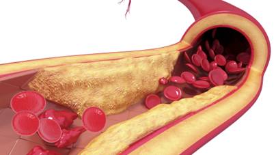
Carotid artery disease involves the narrowing of the carotid arteries in the neck, which supply blood to the brain. This can lead to strokes if left untreated. Doppler ultrasound is vital in diagnosing and managing this condition.
Deep Vein Thrombosis (DVT) involves blood clots forming in deep veins, often in the legs. These clots can be painful and, if they travel to the lungs, can cause a pulmonary embolism. Doppler ultrasound is a key tool in diagnosing DVT.
Varicose veins are swollen, twisted veins that are often visible under the skin. While often seen as a cosmetic issue, they can lead to more serious health problems like ulcers or blood clots. Doppler ultrasound helps evaluate and manage varicose veins.
One of the valuable aspects of Doppler ultrasound is its ability to be used repeatedly without risk. This makes it perfect for ongoing monitoring of vascular health, especially in chronic conditions.
Doppler ultrasound is a versatile and essential tool in diagnosing and managing various vascular diseases. Its ability to provide real-time, detailed information on blood flow and vessel health makes it invaluable in modern cardiovascular care. Patients and healthcare providers alike benefit from the non-invasive, radiation-free nature of Doppler ultrasound, which allows for safe and ongoing monitoring of vascular health.
An abdominal aortic aneurysm (AAA) is a bulge in the main artery that supplies blood to the abdomen, pelvis, and legs. If this bulge bursts, it can cause life-threatening bleeding. Doppler ultrasound helps evaluate and monitor AAAs.
Renal artery stenosis is the narrowing of the arteries that supply blood to the kidneys. This can lead to high blood pressure and kidney damage. Doppler ultrasound is essential in diagnosing and managing this condition.
Chronic venous insufficiency (CVI) occurs when the veins in the legs don’t allow blood to flow back to the heart properly. This can cause swelling, pain, and skin changes. Doppler ultrasound is useful in diagnosing and managing CVI.
Doppler ultrasound evaluates the function of veins and their valves. It helps detect any areas where blood is pooling due to faulty valves. Identifying these issues early allows for treatments to improve blood flow and reduce symptoms.
By using color and spectral Doppler, doctors can measure blood flow patterns in the legs. Abnormal flow patterns can indicate CVI. With this information, doctors can recommend interventions such as compression therapy, lifestyle changes, or surgery to improve vein function and patient comfort.
Role in Pre-Surgical Planning
Doppler ultrasound plays a crucial role in planning for vascular surgeries. It provides essential information that helps surgeons prepare for procedures with greater accuracy.
Before surgery, Doppler ultrasound maps out the structure and function of arteries and veins. This detailed mapping ensures that surgeons know the exact locations and conditions of blood vessels, which helps in planning the surgical approach. Accurate mapping can lead to more successful surgical outcomes.
Doppler ultrasound can assess the risks associated with surgery by evaluating the condition of blood vessels. Identifying issues like blockages or weak vessel walls helps surgeons plan for potential complications. Knowing these risks beforehand helps in making the surgery safer and more effective.
Importance in Emergency Situations
Doppler ultrasound is invaluable in emergency situations, providing quick and accurate information that can guide lifesaving decisions.
In emergencies, such as suspected deep vein thrombosis or arterial occlusion, Doppler ultrasound provides rapid diagnosis. Quick identification of blood clots or blockages allows for immediate treatment, potentially preventing more severe complications.
Doppler ultrasound helps guide immediate interventions, such as administering clot-dissolving medications or planning emergency surgery. Accurate and timely information ensures that patients receive the right treatment promptly.
The role of Doppler ultrasound in diagnosing vascular diseases is extensive and vital. Its ability to provide detailed and real-time images of blood flow and vessel health makes it an essential tool for early detection, monitoring, and planning treatments. This technology enhances patient outcomes by enabling accurate diagnoses and guiding effective interventions for various vascular conditions.
The Vital Role of Doppler Ultrasound in Vascular Health
Doppler ultrasound stands as a cornerstone in the diagnosis and management of vascular diseases. Its ability to provide real-time, detailed images of blood flow and vessel structure makes it indispensable in detecting conditions like peripheral arterial disease, carotid artery disease, deep vein thrombosis, and more. This non-invasive, radiation-free diagnostic tool offers significant advantages in early detection and ongoing monitoring, helping to prevent serious complications and improve patient outcomes.
At Modern Heart and Vascular, we leverage the power of Doppler ultrasound to ensure our patients receive accurate diagnoses and personalized treatment plans. Our commitment to using advanced technology like Doppler ultrasound underlines our dedication to exceptional cardiovascular care.
Take the proactive step towards better vascular health. Schedule a consultation with our cardiovascular practice in Houston today to benefit from expert evaluations and comprehensive care. Your heart and vascular health are paramount—let us help you maintain them. Visit our website or call us to book your appointment. Stay ahead of vascular diseases with Modern Heart and Vascular.

CARDIOVASCULAR CENTERS IN HOUSTON, TEXAS
Modern Heart and Vascular, a preventive cardiology medical practice, has several offices around Houston. We have locations in Humble, Cleveland, The Woodlands, Katy, and Livingston.
We are Modern Heart and Vascular Institute, a diagnostic and preventative medicine cardiology practice.
Every heart has a story… What’s yours?
Book an Appointment Today
At the Modern Heart and Vascular Institute, we offer state-of-the-art cardiovascular care with innovative diagnostic tools and compassionate patient care. Our priority at Modern Heart and Vascular Institute is prevention. We help patients lead healthier lives by avoiding unnecessary procedures and surgeries.
Contact us online to learn more and book an appointment. If you’d like to learn more about our practice, read our providers’ bios.
This article does not provide medical advice. It is intended for informational purposes only. It is not a substitute for professional medical advice, diagnosis, or treatment. If you need cardiovascular care, please call us at 832-644-8930.
Every heart has a story…What’s yours?
Choose your appointment at one of our 7 locations